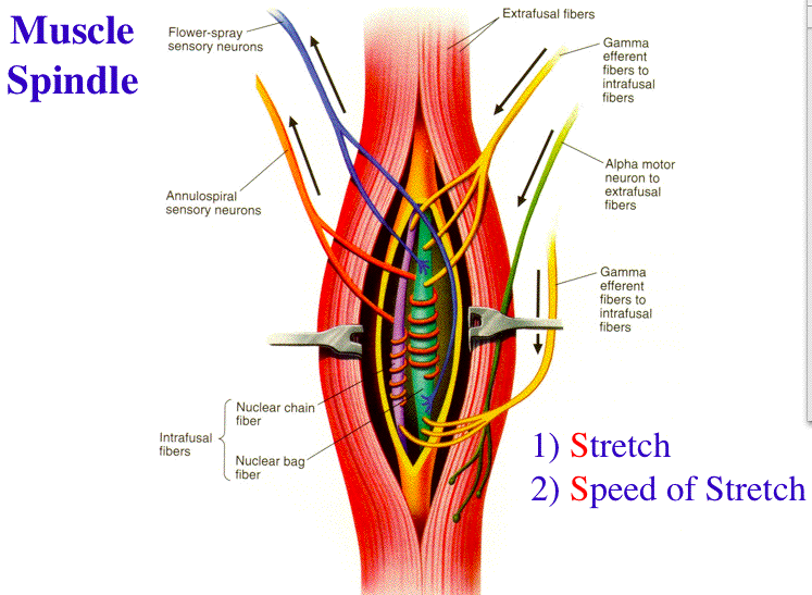

As in the real muscle spindle, the spindle model, under the modulation of γ efferents, responds to the extrafusal muscle fiber length.

The model contains three intrafusal fibers (bag1, bag2, and chain), two efferents (dynamic γ efferent to the bag1 fiber and static γ efferent to bag2 and chain fibers), and two afferents. The smooth muscle cells that can react individually are responsible for the faster smooth muscle reactions, such as the pupil of the eye rapidly shrinking in diameter to protect bright light from dazzling you.A model of the muscle spindle was developed based on its anatomical structure. The way signals move among groups of smooth muscle cells is via junctions between the cells called "gap junctions." Typically, the cells that communicate with one another tend to produce long and slow contractions, such as in the digestive tract. The cells that can contract on their own do not need to communicate with one another, but other cells that contract as a group do need to be able to communicate. These swellings release nerve signals that interact directly with the nearby cell to increase or decrease contraction. Instead, the nerve endings have little swellings on their ends that are near the smooth muscle cell. Smooth muscle cells do not have a special junction where the contraction impulse from a nerve ending attaches. This allows the sheet of muscle that contains them to wrap tightly and evenly around a hollow center of an artery, for example. This lattice shape means that as the cells contract, they get smaller and less elongated in all directions. Instead, the actin and myosin are arranged in a lattice shape throughout the cell. According to "Wheater's Functional Histology," smooth muscle does not have myofibrils. When you can see stripes under the microscope, it means the actin and myosin are arranged in a specific, regular pattern called myofibrils, which appear as stripes. This is why this type of muscle is called "smooth." All muscles need a skeleton of fibers that can contract and relax, and in all muscle types, proteins called actin and myosin form this skeleton. Unlike other types of muscle, smooth muscle does not have a stripy appearance, or striations, when viewed under the microscope. Golgi apparatus, where proteins are modified, are also in this area, along with polyribosomes, which build proteins. Examples of organelles found in the center of the cell include mitochondria - the energy powerhouses of the cells - and rough endoplasmic reticulum, where newly formed proteins are stored. In the vicinity of the nucleus are various organelles, which are individual structures with specific cell functions. The nucleus of a smooth muscle cell, which contains the DNA, is located in the center of the cell, where the cell is widest. For example, when smooth muscle sheets surround an artery, contraction makes the interior of the artery smaller, and relaxation helps the artery dilate. The cells typically form a sheet wrapping around a hollow center when they contract, they make the hollow center constrict. Some smooth muscle cells contract individually, whereas others contract as a group. Some of the cells have ends that are split in two. As smooth muscle cells have an elongated shape, they can fit together in an arrangement in which the middle portion, which is the widest, tucks in next to the thin end of the neighboring cells.


 0 kommentar(er)
